

"Played out"; Boy ditches video violent games
Cantore Arithmetic is able to state;
a.
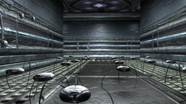
b. I understand words YOUR MACHINE as a backwards Tarantula I just didn’t know exactly what word Anthropoid it was so, remember the Bug in the Film is great and in the word understanding of the Tarantula the Bug in the Film is able to be word Machined so word Built it just needs to go word Backwards from the actual Tarantula so the Mechanical is pulling the opposite word direction from the Tarantula! Here is a good word START as understanding the equated word to show the line just change the word can and then just build as these Cans will show you that the Tarantula has his head going the correct direction and the other Can is the Front End of words YOUR MACHINE!!!!!!

8Arthropoda: Arachnida
Benjamin Kennedy1, Steven A. Trim2, Damien Laudier3, Elise E.B. LaDouceur4, and John E. Cooper5
1 Veterinary Invertebrate Society, Venomtech Ltd, Discovery Park, Sandwich, Kent, UK
2 Venomtech Ltd, Discovery Park, Sandwich, Kent, UK
3 Laudier Histology, New York, NY, USA
8.1 Introduction
8.1.1 Taxonomy
8.1.2 Life History
The life expectancy of arachnids ranges from a few weeks (some species of mites) to several decades, with one mygalomorph spider reported to have lived to 43 years (Mason et al. 2018). Females often outlive males. Arachnids typically lay eggs except for Scorpiones, which develop eggs that hatch internally. In oviparous species, sometimes eggs are merely dropped into the environment, but in many arachnids, there is parental care, in the form of protective webs (some spiders). Mothers may transport egg‐cases or young (some spiders and scorpions). Arachnids undergo an “incomplete metamorphosis”; a juvenile that resembles the adult emerges from the egg and then, through a series of skin changes (ecdysis), grows to maturity. Arachnids vary greatly in their methods of feeding. Some mites feed on plant juices, others on organic debris. Most ticks ingest blood or hemolymph. The majority of spiders and scorpions are “carnivorous”; the former ingest prey through external digestion of internal organs and hemolymph.
8.1.3 Relevance for Conservation, Agriculture, Trade, Etc.
Some arachnids are threatened species, either at an international level (through Convention on Trade in Endangered Species [CITES] and the IUCN) or nationally (species and/or habitat protection). A substantial number of arachnids, notably ticks and mites, are a threat to agriculture (livestock/crops). Of the venomous arachnids (the majority of spiders, all scorpions, some pseudoscorpions), a few are hazardous to humans and other vertebrate animals, some are even lethal. Some species, such as harvestmen, are harmless. Trade in arachnids is restricted to species that are kept in zoos or by private arachnologists, and those that are used in biomedical research. The list of traded species is constantly growing with new species turning up in the pet trade before scientific description, as evidenced by taxonomic papers citing captive material as their source, such as for some Tapinauchenius (Hüsser 2018). Traded species are predominantly theraphosid spiders, but many scorpions and true spiders are also frequently traded in the international pet trade, although few are protected. As of January 2019, only 26 species are listed under the CITES all Appendix II, EU Annex B. These are species in the genus Brachypelma (family Theraphosidae), Aphonopelma albiceps, and Aphonopelma pallidum (family Theraphosidae, previously in genus Brachypelma), and four species of Scorpionidae of the genus Pandinus (Pandinus dictator, Pandinus gambiensis, Pandinus imperator, and Pandinus roeseli) (UNEP 2019). In total, 286 species have been evaluated by the IUCN redlist (www.IUCNredlist.org) with 52 species found to be critically endangered, thus conservation evaluation in the arachnid taxa to date has only evaluated a small percentage of the global species and these are heavily biased to the large spiders.
In biomedical research, peptides found in the venom of arachnids are becoming increasingly important in studying drug targets such as ion channels and others (Escoubas and Rash 2004). Although pain and neuroscience are the predominant indications (Trim and Trim 2013), other indications are also pursued including pesticides (Hardy et al. 2013). Alpha‐latrotoxin (Tzeng and Siekevitz 1978) from the black widow spider (Latrodectus mactans) and imperatoxin from the emperor scorpion (Pandinus imperator) have been used to understand the biology of neurons.
Zoonosis is, strictly, an infection that can be acquired by humans from a vertebrate animal but it should be noted that certain arachnids can be a source of nematodes of the family Panagrolaimidae (Pizzi 2009; Pizzi et al. 2003), which can be of medical concern to humans.
8.2 Gross Anatomy
Although all arthropods have abundant segmentation embryonically, arachnids have reduced outward segmentation in adults. This is most pronounced in animals from the subclass Acari, which have no external segmentation. Most other arachnids are segmented into an anterior prosoma and a posterior opisthosoma with a narrow waist called a pedicel (the seventh embryonic segment) joining the two. The opisthosoma is segmented in eurypterids and scorpions; most other arachnids do not have opisthosomal segmentation. Some species also have a telson, or tail. In scorpions, the telson contains a venom gland and is connected to the aculeus (stinger), and in palpigrades, uropygids, and schizomids it acts as a sensory flagellum.
Arachnids have six paired appendages. The first are the chelicerae, the second are the pedipalps, and the last four are the walking legs. Each chelicera is attached to the cranial aspect of the prosoma via a fixed basal segment that is articulated with a movable fang. The fangs typically rest in a groove in the basal segment, from which they pivot outward to strike prey. Venom is delivered through a slit in the subterminal portion of the fang. The chelicerae are not only used for capturing prey, but also for defense and for grasping various objects. These are most well developed in the Solifugidae (Klann 2009). The second appendages, the pedipalps, have segmentation similar to the legs except they lack one segment, the metatarsus. Pedipalps are not used for locomotion (except in Aranae), but for capturing and manipulating prey. The first segment of the pedipalps, the coxa, forms the preoral cavity and is frequently modified to macerate prey. In all but the Aranae, Solifugidae and Ricinulei, the pedipalps form grasping claws with a single mobile finger opposing a fixed finger. Additionally, male spiders produce a bulbous distension of the terminal aspect of the pedipalps, called the palpal bulb, upon sexual maturity, for copulation. Four pairs of walking legs attach to the prosoma caudal to the pedipalps. Walking legs have seven segments, including (from proximal to distal) the coxa, trochanter, femur, patella, tibia, metatarsus, and tarsus. The tip of the tarsus bears claws. The number of leg segments and number of claws may vary between species (Ruppert et al. 2004).
Paired eyes are on the craniodorsal aspect of the prosoma; in Araneidae, there are four pairs on an ocular tubercle. The cranioventral aspect of the opisthosoma has a number of book lung slits (in pulmonate species) that are adjacent to the epigastric furrow. The cloaca and spinnerets are located on the caudoventral aspect of the opisthosoma. The opisthosoma is typically covered by setae in Theraphosidae. Setae are detachable and urticating in most new‐world theraphosids. There is a great deal of variation of the gross anatomy within Arachnida. Gross external anatomic features are provided for Theraphosidae in Figure 8.1, Scorpiones in Figures 8.2 and 8.3, and Acaridia in Figure 8.4.
8.2.1 Dissection
There are multiple dissection techniques that can be applied to arachnids. The method described here is the authors’ preferred technique with respect to Araneae. The dissection method may need to be modified for other orders of Arachnida. Successful microdissection is greatly aided by stereo microscopy with 2×–5× magnification and through the use of size 5 microforceps and bow scissors.
The prosoma and opisthosoma are dissected separately starting with the prosoma. The prosomal carapace is cut at its periphery with shears and then gently lifted to expose the underlying muscle and nerves (Figure 8.5a,b). Muscular attachments from the carapace to the underlying structures will need to be cut for the carapace to be easily removed. The eyes and the optic stalk can be visualized and these can be followed down to the cranial part of the supraesophageal ganglion (sometimes referred to as the central ganglion, protocerebrum or “brain”) (Figure 8.6).
Gentle dissection of dorsal musculature will expose a hard cartilaginous structure, the endosternite. In Araneae, the endosternite is centrally located and proximal to the pedicle originating from the dorsal opisthosoma. Dorsal to the endosternite is the esophagus and stomach which leads to the opisthosoma. Beneath the endosternite is the central ganglion. The central ganglion can also be easily identified by removing a leg and following the nerve branch to the central ganglion.

Figure 8.1 Gross anatomy. Dorsal (left) and ventral (right) view of theraphosid spider.


No comments:
Post a Comment